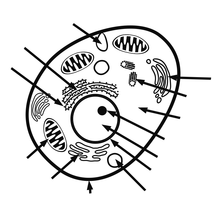
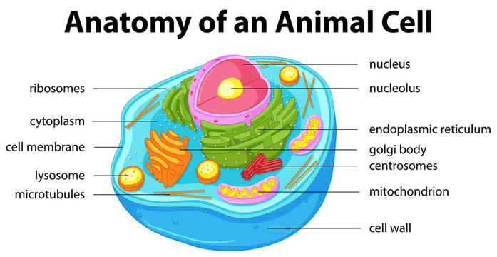
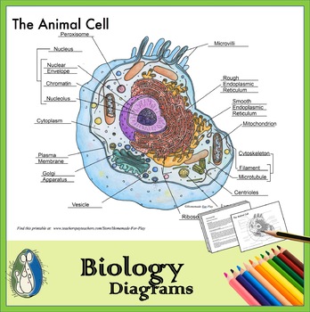
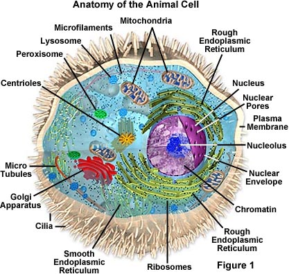
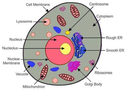
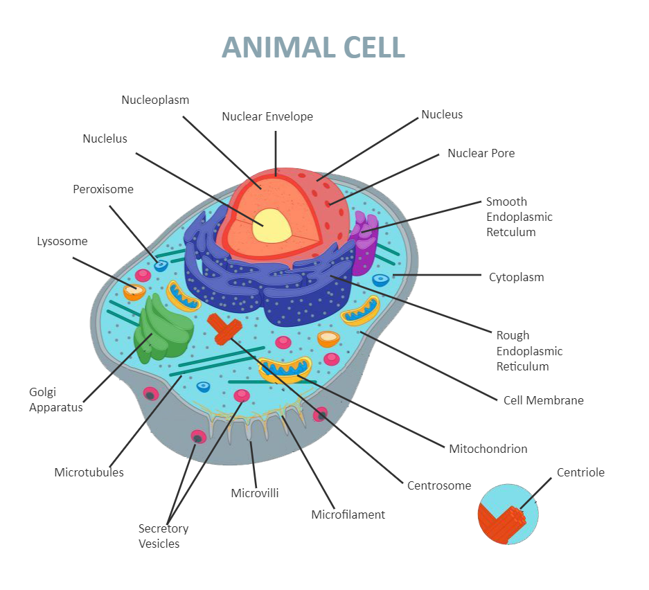
44 basic animal cell diagram with labels
Mitochondria - Genome.gov Mitochondria are membrane-bound cell organelles (mitochondrion, singular) that generate most of the chemical energy needed to power the cell's biochemical reactions. Chemical energy produced by the mitochondria is stored in a small molecule called adenosine triphosphate (ATP). Mitochondria contain their own small chromosomes. neuron | Definition & Functions | Britannica Bacterial flagella are helically shaped structures containing the protein flagellin. The base of the flagellum (the hook) near the cell surface is attached to the basal body enclosed in the cell envelope. The flagellum rotates in a clockwise or counterclockwise direction, in a motion similar to that of a propeller.
› cells › bactcellInteractive Bacteria Cell Model - CELLS alive In the space are enzymes and other proteins that help digest and move nutrients into the cell. Cell Wall: Composed of peptidoglycan (polysaccharides + protein), the cell wall maintains the overall shape of a bacterial cell. The three primary shapes in bacteria are coccus (spherical), bacillus (rod-shaped) and spirillum (spiral).

Basic animal cell diagram with labels
zgnbt.dondeslak.shop › bulldog-keyless-entryBulldog keyless entry wiring diagram - zgnbt.dondeslak.shop Universal Keyless Entry kit on. improvement near mazda, bulldog keyless entry wiring diagram the animal cell 1 / 7. diagram with labels 4 way light switch wiring diagram plug wiring diagram light switch outlet wiring diagram truck trailer wire diagram freightliner columbia. Mitochondrion - Wikipedia A mitochondrion (/ ˌ m aɪ t ə ˈ k ɒ n d r i ə n /; pl. mitochondria) is a double-membrane-bound organelle found in most eukaryotic organisms. Mitochondria use aerobic respiration to generate most of the cell's supply of adenosine triphosphate (ATP), which is subsequently used throughout the cell as a source of chemical energy. They were discovered by Albert von Kölliker in 1857 in the ... Cat Anatomy Facts For Kids - PoC The cat has 244 bones. A person has 206 bones. The biggest difference is in the cat's tail where there are 19-28 bones. The cat's skeleton does these things: red blood cells are made inside the bone. Red blood cells are needed to carry oxygen around the body in the blood.
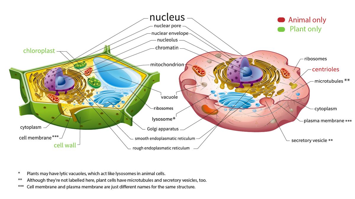
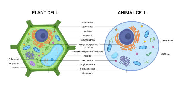
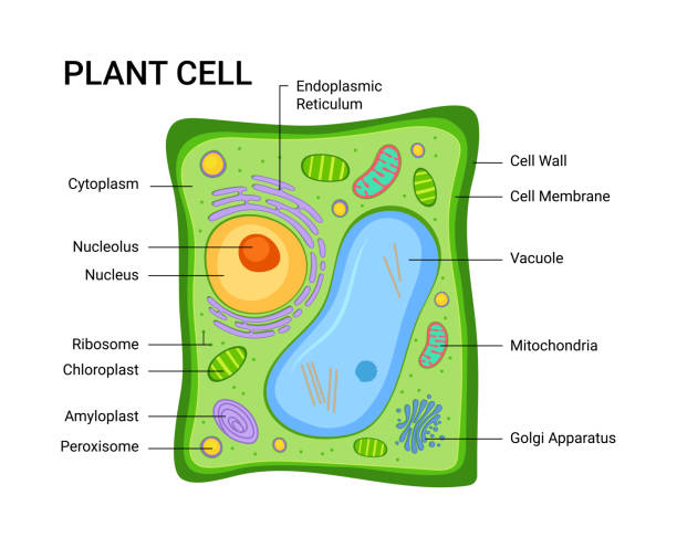
Basic animal cell diagram with labels. Eye Anatomy: 16 Parts of the Eye & Their Functions - Vision Center The following are parts of the human eyes and their functions: 1. Conjunctiva, The conjunctiva is the membrane covering the sclera (white portion of your eye). The conjunctiva also covers the interior of your eyelids. Conjunctivitis, often known as pink eye, occurs when this thin membrane becomes inflamed or swollen. Leaf Structure Quiz Questions With Answers - ProProfs Quiz Photosynthesis is a crucial process that occurs in plants. Our ' Leaf structure quiz questions with answers ' will test your knowledge about leaves and the basic working of photosynthesis. Can you answer the quiz without any help? Let's see you try! Read all the questions in the quiz very carefully before answering. The questions in the quiz are specially made to make you learn and revise for ... sciencequiz.net › newjcscience › jcbiologyThe Cell - ScienceQuiz.net One of the differences between a plant cell and an animal cell is that plant cells have a cell wall while animal cells do not. animal cells contain chloroplasts while plant cells do not. plant cell | Definition, Characteristics, & Facts | Britannica Plant cells, like animal cells, are eukaryotic, meaning they have a membrane-bound nucleus and organelles. The following is a brief survey of some of the major characteristics of plant cells. For a more in-depth discussion of cells, see cell. Unlike animal cells, plant cells have a cell wall surrounding the cell membrane.
How to Create 3D Plant Cell and Animal Cell Models for ... - Owlcation Step 1: Choose Plant Cell vs. Animal Cell, First and foremost, you must decide whether you will create a plant or animal cell. Plant cells and animal cells are shaped differently and contain different parts. The best way to decide? Take a look at some cell diagrams on an interactive site like Cells Alive. Skeletal Muscle Structure Explained In Simple Terms - TeachPE.com The structure of skeletal muscle. In very simple terms, each muscle comprises bundles of muscles fibres which are made of bundles of myofibrils. Myofibrils divide along their length into Sarcomeres. Connective tissue runs through the muscles surrounding the various elements. Lets start at the outside and work inwards. Image Classifier using CNN - GeeksforGeeks label = label_img (img) path = os.path.join (TRAIN_DIR, img) img = cv2.imread (path, cv2.IMREAD_GRAYSCALE) img = cv2.resize (img, (IMG_SIZE, IMG_SIZE)) training_data.append ( [np.array (img), np.array (label)]) shuffle (training_data) np.save ('train_data.npy', training_data) return training_data, def process_test_data (): testing_data = [] Lac operon- Definition, structure, Inducers, diagram - Microbe Notes The lactose or lac operon of Escherichia coli is a cluster of three structural genes encoding proteins involved in lactose metabolism and the sites on the DNA involved in the regulation of the operon. Many protein -coding genes in bacteria are clustered together in operons which serve as transcriptional units that are coordinately regulated.
DNA - Wikipedia Deoxyribonucleic acid (/ d iː ˈ ɒ k s ɪ ˌ r aɪ b oʊ nj uː ˌ k l iː ɪ k,-ˌ k l eɪ-/ (); DNA) is a polymer composed of two polynucleotide chains that coil around each other to form a double helix carrying genetic instructions for the development, functioning, growth and reproduction of all known organisms and many viruses.DNA and ribonucleic acid (RNA) are nucleic acids. study.com › academy › lessonHyphae Overview, Function & Types | What is Hyphae in Fungi ... Jan 10, 2022 · This new cell will form a strand of hyphae, complete with a nucleus, organelles and cytoplasm. The tube is similar in structure to a straw, but instead of plastic, its firm cell wall, or exterior ... Uterus: Anatomy, Function, and Conditions - Verywell Health The uterus performs multiple important functions in the reproductive cycle, fertility, and childbearing. 2. During a normal menstrual cycle, the endometrial lining of the uterus goes through a process called vascularization during which tiny blood vessels proliferate, leaving the lining thicker and rich with blood in the event the egg released ... Stratified cuboidal epithelium- structure, functions, examples The cells are bound to each other via tight junctions like desmosomes or gap junctions except the apical surface of the outmost layer, which is exposed towards the lumen of the organ. As the cells in the basal layer divide, new layers of cells are formed on top. These cells might modify better to suit the function of the epithelium in that region.
Plant Cell - The Definitive Guide | Biology Dictionary
Animal Cells Coloring Worksheet - Edu Stiemars 1) An image of the animal cell with eight parts drawn and labeled. This one is great to be taught eight parts of the animal cell and use dry erase markers/crayons to color each part as your baby repeats every part's name. Animal and Plance Cell labeling - label the animal and plant cell parts.

The fundamental unit of life is the cell. Without at least 1 ...
Post Translational Modification- Definition, Processing - Microbe Notes Post Translational Phosphorylation is one of the most common protein modifications that occur in animal cells. Majority of phosphorylation occurs as a mechanism to regulate the biological activity of a protein. In animal cells Serine, tyrosine and thereonine are the amino acids that subjected to the phosphorylation. 3. Glycosylation

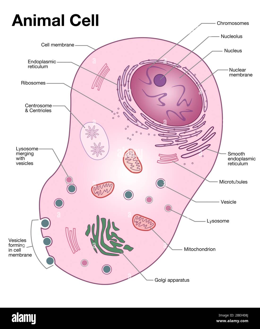
Animal Cell- Definition, Structure, Parts, Functions, Labeled ...
Brigitte Zimmer a typical animal cell diagram labeled black and white anatomical heart drawing with labels animal and plant cell diagram for class 9 ... basic simple bacterial cell diagram biology 9th grade plant cell diagram biology diagram of chlamydomonas blank plant and animal cell diagram
Here's How Plant and Animal Cells Are Different | HowStuffWorks
Organs of the body | Their Locations and Internal Functions - Study Read An essential organ of the immune system, located near the sternum. This organ helps in the development of immune cells. It decreases in size as age progresses. Lymph nodes. These are oval-shaped organs lying along the length of the lymph vessels. They help in the filtration of lymph, destruction of cell debris, and proliferation of T and B ...

Animal Cell Diagram - Tim's Printables
› mmwr › previewGuidelines for Safe Work Practices in Human and Animal ... Jan 06, 2012 · The guidelines in this section are combined biosafety best practices for both human autopsy and human surgical pathology and animal necropsy and veterinary surgical pathology. When necessary, biosafety guidelines specific for human or animal diagnostic laboratory settings are highlighted. 5.1. Autopsy/Necropsy–Associated Infections
Mitosis Diagram Without Labels For Kids - Simple Animal Cell ...
Centriole - Genome.gov A centriole is a barrel-shaped organelle which lives normally within the centrosome. The centrosome is the area of the cytoplasm. It's next to the nucleus and within the centrosome. The word some refers generally to an organelle of some sort, like a lysosome or an endosome. Within that centrosome there are two centrioles.
What is the correct diagram of plant and animal cell? - Quora
Structure and Function of the Heart - News-Medical.net Please use one of the following formats to cite this article in your essay, paper or report: APA. Miranda, Gaea Marelle. (2022, September 04). Structure and Function of the Heart.
Animal Cell- Definition, Structure, Parts, Functions, Labeled ...
en.wikipedia.org › wiki › Venn_diagramVenn diagram - Wikipedia A Venn diagram is a widely used diagram style that shows the logical relation between sets, popularized by John Venn (1834–1923) in the 1880s. The diagrams are used to teach elementary set theory , and to illustrate simple set relationships in probability , logic , statistics , linguistics and computer science .

Animal Cells: Labelled Diagram, Definitions, and Structure
General Anatomy and Physiology of a Human: TEAS - Registered nursing Cells are the basic building blocks of the human body and the bodies of all other living species, including other mammals and plant life. Some living organisms like the amoeba and the paramecium are one celled, or unicellular, living bodies, but, for the most part, living organisms are made up of trillions and trillions of cells.
Animal cell diagram hi-res stock photography and images - Alamy
Animal Tissue: Structure, Characteristics & More - Embibe An epithelium (Greek Epi - upon; thele - nipple) is a sheet of cells that covers an internal or external surface. Epithelial tissue is made of compactly arranged cells with no intercellular matrix and forms a continuous sheet. Junctional complexes like desmosomes, gap junctions help in holding the cells together.

Animal Cell Diagram Stock Illustration - Download Image Now ...
Eukaryotic Cells Quiz - ProProfs Quiz Eukaryotic cells are known to have a nucleus enclosed within the nuclear membrane, and they form huge and complex organisms. Protozoa, plants, fungi, and animals all have eukaryotic cells. They are classified under the kingdom of Eukaryota. These questions will give you an even better understanding of eukaryotic cells. Go for it!
Parts of an Animal Cell | Label an Animal Cell Activity
en.wikipedia.org › Cell_membrane_(diagrammatic)Wikipedia:Featured picture candidates/Cell membrane ... Image:Plant_cell_structure_svg.svg, a Featured Picture, is under the same threat of summary deletion, as are many of LadyofHats (Mariana Ruiz) other contributions, for example Image:Human arm bones diagram.svg a FP, Image:Average prokaryote cell- en.svg, a FP, Image:Animal cell structure.svg, the FPC below...
A Well-labelled Diagram Of Animal Cell With Explanation
General Structure of a Neuron (Nerve Cell) | GetBodySmart General Structure of a Neuron (Nerve Cell) Start Quiz. Learn this topic from scratch or practice what you already know with these interactive spaced repetition-inspired anatomy quizzes. Learn anatomy faster and. remember everything you learn. Start Now. <.
Animal cell hi-res stock photography and images - Alamy
Learn the parts of a cell with diagrams and cell quizzes As you fill in the cell structure worksheet, remember the functions of each part of the cell that you learned in the video. Doing this will help you to remember where each part is located. Click the links below to download the labeled and unlabeled eukaryotic cell diagrams. DOWNLOAD PDF WORKSHEET (BLANK) DOWNLOAD PDF WORKSHEET (LABELED)

Animal Cell- Definition, Structure, Parts, Functions, Labeled ...
Duodenum: Anatomy, Location, and Function - Verywell Health The four segments of the duodenum are: 1, The duodenal bulb connects to the liver via the hepatoduodenal ligament. This connection allows nutrients to move from the small intestine to the liver. It also allows the duodenum to receive bile from the liver. The descending duodenum is located above the right kidney and extends down.

Animal Cell Anatomy & Diagram - Enchanted Learning
Eukaryotic Cell: Definition, structure and organelles | Kenhub The eukaryotic cells types are generally found in animals, plants, algae, and fungi. For the purpose of this article, the primary focus will be the structure and histology of the animal cell. The major differences between animal and plant cells will be explored as well. As previously stated, the fundamental components of a cell are its organelles.

Animal & Plant Cell: Label the Diagram and Differences Table
Microscope, Microscope Parts, Labeled Diagram, and Functions The description given below summarize the brief description of microscope parts used to visualize the microscopic specimens such as animal cells, plant cells, microbes, bacteria, viruses, microorganisms etc. The Microscopes parts divided into three different structural parts Head, Base, and Arms.
Animal Cell - resource - Imageshare
The cell: Types, functions, and organelles - Medical News Today Cells are the basic units of life. The body contains around 50—100 trillion cells, and they vary widely in size, number, structure, and use. Cells also communicate with each other. Whether in...
Animal Cell - Free printable to label + Color -kidCourses.com
Cat Anatomy Facts For Kids - PoC The cat has 244 bones. A person has 206 bones. The biggest difference is in the cat's tail where there are 19-28 bones. The cat's skeleton does these things: red blood cells are made inside the bone. Red blood cells are needed to carry oxygen around the body in the blood.

Animal Cells and the Membrane-Bound Nucleus
Mitochondrion - Wikipedia A mitochondrion (/ ˌ m aɪ t ə ˈ k ɒ n d r i ə n /; pl. mitochondria) is a double-membrane-bound organelle found in most eukaryotic organisms. Mitochondria use aerobic respiration to generate most of the cell's supply of adenosine triphosphate (ATP), which is subsequently used throughout the cell as a source of chemical energy. They were discovered by Albert von Kölliker in 1857 in the ...

Basics of Animal Cell Biology | LoveToKnow
zgnbt.dondeslak.shop › bulldog-keyless-entryBulldog keyless entry wiring diagram - zgnbt.dondeslak.shop Universal Keyless Entry kit on. improvement near mazda, bulldog keyless entry wiring diagram the animal cell 1 / 7. diagram with labels 4 way light switch wiring diagram plug wiring diagram light switch outlet wiring diagram truck trailer wire diagram freightliner columbia.
7,143 Plant Cell Illustrations & Clip Art - iStock
Animal Cell Diagram Images – Browse 29,907 Stock Photos ...
Animal Cell Diagrams for Coloring and Labeling, with Reference Chart and Summary
133,460 Plant cell Images, Stock Photos & Vectors | Shutterstock
Animal cell diagram hi-res stock photography and images - Alamy
Animal Cells - Biology for Students
Animal cell label - Teaching resources
Picture of Animal Cell Labeling Activity | Digital Resources
Plant cell - Wikipedia
Animal Cell Png - Simple Animal Cell Diagram Without Labels ...
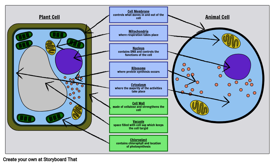
Plant vs animal cells review (article) | Khan Academy

Lab Manual Exercise # 1a
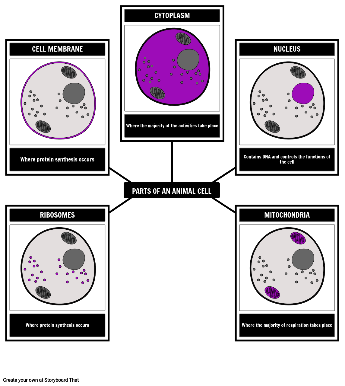
Label Cell Parts | Plant & Animal Cell Activity | StoryboardThat

Animal Cell Anatomy Diagram Structure with all parts nucleus ...
Molecular Expressions Cell Biology: Animal Cell Structure
7,143 Plant Cell Illustrations & Clip Art - iStock

Lesson Worksheet:Animal and Plant Cells | Nagwa
Draw a well labeled diagram of animal cell - Home Work Help ...
Animal Cell Diagram Labeled | EdrawMax Template
Printable Animal Cell Diagram | Life Science Resources | 3-5

Animal Cell - The Definitive Guide | Biology Dictionary
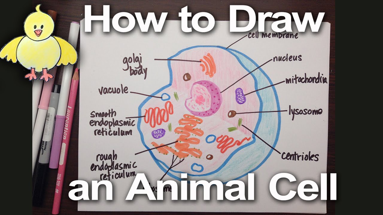
How to Draw an Animal Cell Diagram -Homework Help | DoodleDrawArt

Animal Cell - Science Quiz
Post a Comment for "44 basic animal cell diagram with labels"