39 fluorescent labels and light microscopy
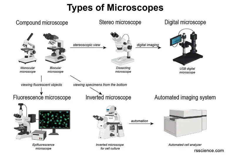
Fluorescence Microscopy vs. Light Microscopy - News Medical Nov 27, 2018 ... Light microscopy does much what the name implies: visible light and magnifying lenses are used to view small objects. Light microscopes are the ... Geminate labels programmed by two-tone microdroplets ... - Nature Jan 29, 2021 · Guo and Li et al. recently have reported an interesting dual-mode CLC system induced by light-driven fluorescent chiral switches, in which the reflection wavelength and fluorescence intensity were ...
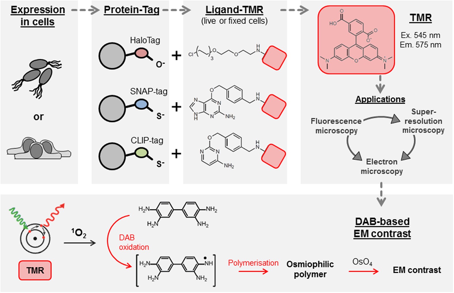
Genetically encoded fluorescent tags | Molecular Biology of the Cell Oct 13, 2017 ... These tags have revolutionized cell biology by allowing nearly any protein to be imaged by light microscopy at submicrometer spatial ...

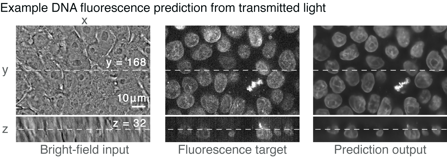
Fluorescent labels and light microscopy
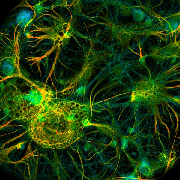
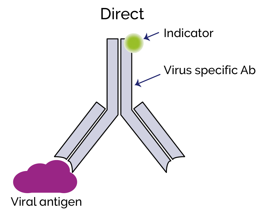
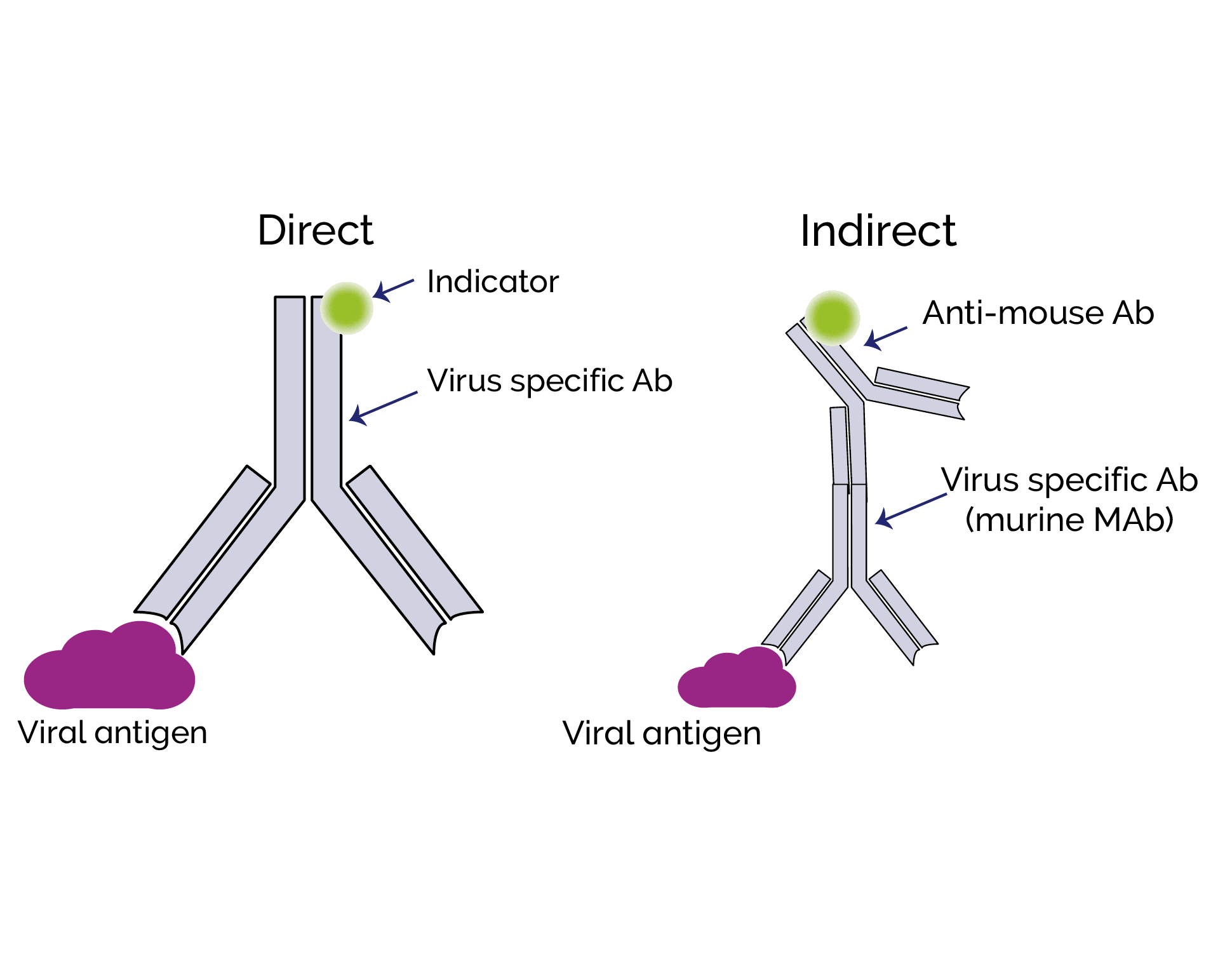
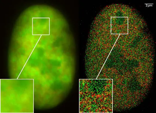
Fluorescent Labeling - What You Should Know - PromoCell Fluorescent labeling is the process of binding fluorescent dyes to functional groups contained in biomolecules so that they can be visualized by fluorescence ... Fluorescence microscope - Wikipedia A fluorescence microscope is an optical microscope that uses fluorescence instead of, or in addition to, scattering, reflection, and attenuation or ... Super-resolution microscopy - Wikipedia Integrated correlative light and electron microscopy. Combining a super-resolution microscope with an electron microscope enables the visualization of contextual information, with the labelling provided by fluorescence markers. This overcomes the problem of the black backdrop that the researcher is left with when using only a light microscope.
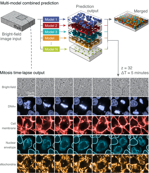
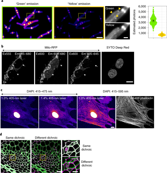
Fluorescent labels and light microscopy. Bright light, better labels | Nature Oct 5, 2011 ... Attaching light-emitting labels to a protein can reveal when and where in a cell it functions, but usually the details are fuzzy. Optical ... [TALK 3] Fluorescent Labelling and Light Sheet Microscopy - YouTube Jan 31, 2022 ... Fluorescent Labelling and Light Sheet Microscopy Speaker: Ben Sutcliffe, MRC Laboratory of Molecular Biology, UKThe LMB Light Microscopy ... Recent Advances in Fluorescent Labeling Techniques for ... - NCBI The fluorescent-labeled probes hybridize with the complementary RNA or DNA molecules in the cells and are used to detect the particular gene expression in the ... Fluorescence - Wikipedia A perceptible example of fluorescence occurs when the absorbed radiation is in the ultraviolet region of the electromagnetic spectrum (invisible to the human eye), while the emitted light is in the visible region; this gives the fluorescent substance a distinct color that can only be seen when the substance has been exposed to UV light ...
Fluorescent Labelling, FRET & Light Sheet Microscopy – Ben Sutcliffe Jan 27, 2021 ... The LMB Light Microscopy Facility supports a diverse range of light microscopy imaging techniques within the LMB including single molecule ... Förster resonance energy transfer - Wikipedia In fluorescence microscopy, fluorescence confocal laser scanning microscopy, as well as in molecular biology, FRET is a useful tool to quantify molecular dynamics in biophysics and biochemistry, such as protein-protein interactions, protein–DNA interactions, and protein conformational changes. For monitoring the complex formation between two ... ProSciTech Laboratory supplies and Lab equipment for Histology, Pathology, Light Microscopy, Electron Microscopy and specialist researchers. Basics of FRET Microscopy | Nikon’s MicroscopyU The first fluorescent protein biosensor was a calcium indicator named cameleon, constructed by sandwiching the protein calmodulin and the calcium calmodulin-binding domain of myosin light chain kinase (M13 domain) between enhanced blue and green fluorescent proteins (EBFP and EGFP). In the presence of increasing levels of intracellular calcium ...
Click-ExM enables expansion microscopy for all biomolecules Dec 07, 2020 · Expansion microscopy (ExM) allows super-resolution imaging on conventional fluorescence microscopes, but has been limited to proteins and nucleic acids. ... Using 18 clickable labels, we ... Fluorescent Dyes | Science Lab - Leica Microsystems Jan 10, 2022 ... Alexa Fluor® dyes are a big group of negatively charged and hydrophilic fluorescent dyes, frequently used in fluorescence microscopy. All the ... Different Ways to Add Fluorescent Labels | Thermo Fisher Scientific Using fluorescence provides greater contrast compared to viewing your samples with brightfield microscopy alone. Labeling various targets with separate ... Super-resolution microscopy - Wikipedia Integrated correlative light and electron microscopy. Combining a super-resolution microscope with an electron microscope enables the visualization of contextual information, with the labelling provided by fluorescence markers. This overcomes the problem of the black backdrop that the researcher is left with when using only a light microscope.
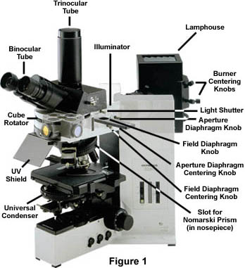
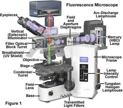
Fluorescence microscope - Wikipedia A fluorescence microscope is an optical microscope that uses fluorescence instead of, or in addition to, scattering, reflection, and attenuation or ...
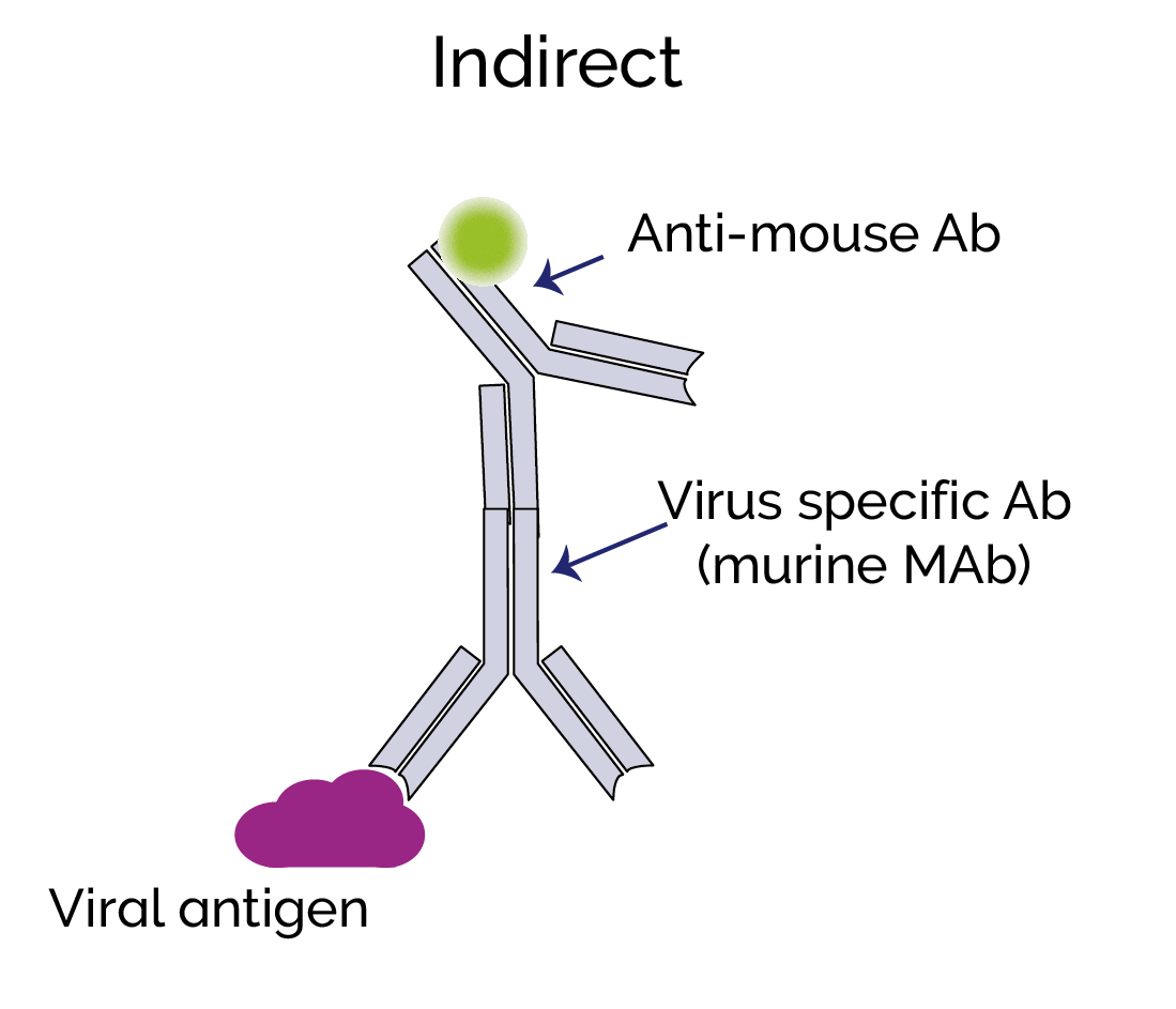
Fluorescent Labeling - What You Should Know - PromoCell Fluorescent labeling is the process of binding fluorescent dyes to functional groups contained in biomolecules so that they can be visualized by fluorescence ...











Post a Comment for "39 fluorescent labels and light microscopy"